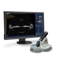
The UBM Plus probe has a guarded tip to prevent damage to both the patient and the probe. The UBM Plus is used for anterior segment imaging. This unit is an incredibly portable device because the 48 MHz probe plugs directly into any Windows™ laptop or desktop computer. This unit features an all-in-one probe design to eliminate signal loss and provide the sharpest images possible.
Features:
High-Definition Imaging - Detailed structure definition including cornea, iris, ciliary body, zonules, crystalline lens, and intraocular lens, as well as pathologies
State-of-the-Art Probe Design - Sharper, more focused images due to the elimination of signal loss
Easy To Set Up Defaults – Smooth customer interface and automatic EMR exports that decrease examination time, increase patient throughput, and increase profit
Unsurpassed Data Analysis - Contains tools for measuring sulcus-to-sulcus, anterior chamber depth, positioning of intraocular lenses, and filtration angle of the eye.
Adjustable video loop.
Fully upgradeable software.
EMR compatible.
DICOM ready.
Multiple measuring velocities for ACD.
PDF reporting.
Fully adjustable gain-control before, during, or after scan.
Data Archive – Network or external location.
Accessories Included: Software installation, USB stick, probe holder, footswitch, wireless mouse, ophthalmic gel, and scleral shell
Weight: 6 oz.
Dimensions: 7” long X 1.25” diameter
Frequency: 48 MHz
Axial Resolution: 0.015mm
Electronic Lateral Resolution: 0.05 mm
Electronic Gain: 0-112 dB
Adjustable Gamma: Linear, S-Curve, Log, Color
Scanning Angle: 30°
Field of View: 32 mm
Frame Rate per Second: 10
Sampling Rate: 2048 (points per line)
Vectors per Frame: 256
Focal Point: 13 mm
Focal Zone: 4 mm
TGC: Yes
Frozen Image Gain Adjustment: Yes
Zoom: 8x Maximum
Reports: Integrated Microsoft Word
Snapshot format: jpeg, bmp, png, tiff, gif
Data archive/ Export capability: Yes
Max. number of frames per scan: 256
Size of cine loop: 16-128 MB
Measurement Calipers with Velocity Adjustment: 4 line, 2 area, 2 angle